
Contrast Sensitivity and Acquired Brain Injury
Patients frequently report decreases in the sharpness of their vision after stroke or other acquired brain injuries. Patients may describe this as a “foggy” blur to their vision. Visual acuity measurements may not always fully explain the patient’s complaints. Contrast sensitivity testing, however, may show that the primary cause is a loss of spatial contrast sensitivity. This can be temporary or permanent. Contrast sensitivity is often a better predictor of functional vision than visual acuity. The real world is made of subtle changes in contrast, not the absolute contrast of black on white as on the visual acuity chart. Additionally, a number of studies have suggested that impaired contrast sensitivity correlates with increased crash risk in driving.
The use of contrast enhancing filters in the light amber-yellow range may be helpful inside and in low contrast situations such as a rainy or foggy day. We find darker amber or blue light blocking filters are often helpful outside. Each patient must be looked at as an individual and it is important to understand that over time the contrast may improve in some patients.
In 1979, we developed our own gradient amber filter (the 508/505) initially for post laser diabetic patients, but found that it was helpful for some acquired brain injury patients. We modify this filter lighter or darker as needed to fit each individual patient. Other contrast enhancing tools we use include the yellow filter overlays we use to mark boundaries for many hemianopsia patients in reading.
It is important that contrast sensitivity testing be added to those tests routinely performed on patients with acquired brain injury, whether of recent origin or post several years.
Contrast Sensitivity Testing
Contrast sensitivity refers to the ability of the visual system to perceive an object or shape from its background. Imagine a grey pen on a slightly darker grey desk. It would be difficult for a patient with poor contrast to distinguish between the two. However, that same patient might easily see a dark letter on a white visual acuity chart because the contrast is very high. Thus contrast sensitivity testing will detect visual impairment that cannot be measured on a visual acuity chart with high contrast black on white.

Contrast sensitivity is a better measurement of real world functioning than visual acuity. Failure to test contrast sensitivity may leave a patient with only moderate visual acuity loss faced with an unrecognized day-to-day visual problems in the real world.
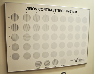
Contrast sensitivity can be tested in many different ways. Some test charts use letters that gradually reduce in contrast. The patient is tested to determine the limit of reducing contrast letters that can be identified. Other contrast sensitivity charts use gratings that gradually change in the contrast and frequency of dark to light oscillations on the plate. Both types can determine the loss of contrast sensitivity.
Poor contrast sensitivity is common in many visual disorders including cataract, diabetic retinopathy and many retinal disorders. We can aid contrast  sensitivity in these patients with the use of high contrast filters, better lighting and by the use of bolder higher contrast materials. In cataract patients, surgical removal of the cataract may significantly improve contrast sensitivity.
The Pelli-Robson Contrast Sensitivity Chart has proven to be a quick and reliable test.
Please contact us if you have any questions.
The Low Vision Centers of Indiana
Richard L. Windsor, O.D., F.A.A.O., D.P.N.A.P.
Craig A. Ford, O.D., F.A.A.O.
Laura K. Windsor, O.D., F.A.A.O.
Ali E. Prible, O.D.
Indianapolis (317) 844-0919
Fort Wayne (260) 432-0575
Hartford City (765) 348-2020